pleural effusion cat ultrasound
Abdominal ultrasounds were performed in 70 cats with pleural effusion and revealed concurrent abdominal effusion in 59 of these cats. The aims of ultrasound guided assessment of pleural effusion are.

Radiographs Felinepleuraleffusionlabeled Vet Medicine X Ray Veterinary Technician
Caudal is to the left of the image.
. The trocar technique is faster and easier. Cats with pleural effusion often have severe respiratory compromise at presentation. The ultrasound US examination was performed less than 6 h after the diagnostic CT scan.
Ultrasound-guided pleural effusion drainage by catheter insertion is a safe and effective procedure. Approximately 1 million people develop this abnormality each year in the United States. To characterize the effusion noting echogenicity of the fluid any loculations solid masses and pleural disease.
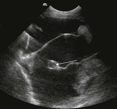
Pleural effusion is present in both hemithoraces e. Cats presenting with pleural effusion are nearly always in respiratory distress ranging from an increased respiratory rate and effort to open mouth breathing. Although the application of ultrasound on the thorax can be limited by aerated lung in normal individuals acoustic window is created with the presence of pleural effusion and non-aerated lung in diseased patients 14For dogs and.
Four standard effusion types recognized in addition to blood. This can be caused by thoracic lymphangiectasia swollen lymph vessels that leak chyle into the pleural space congestive heart failure obstruction of the cranial vena cava the major vein that returns blood to the heart from the front of the body cancer fungal infection feline heartworm. 91 Pediatric abdomen and retroperitoneum 92 Pediatric urinary tract 93 Pediatric scrotum 94 Pediatric gynaecological pathology and infant breast 95 Pediatric head and neck 96 Neonatal brain and spine 97 Infant hip and knee 98 Pediatric thorax.
Abdominal abnormalities identified on ultrasound included abdominal masses lymphadenopathy hepatic venous congestion hepatomegaly splenomegaly renal enlargement small intestinal wall thickening steatitis and pancreatitis. A sample of pleural fluid obtained by piercing the cats chest cavity with a needle will be sent to the laboratory for analysis. Ultrasound has become a common diagnostic procedure in small animal clinical medicine over the past decades.
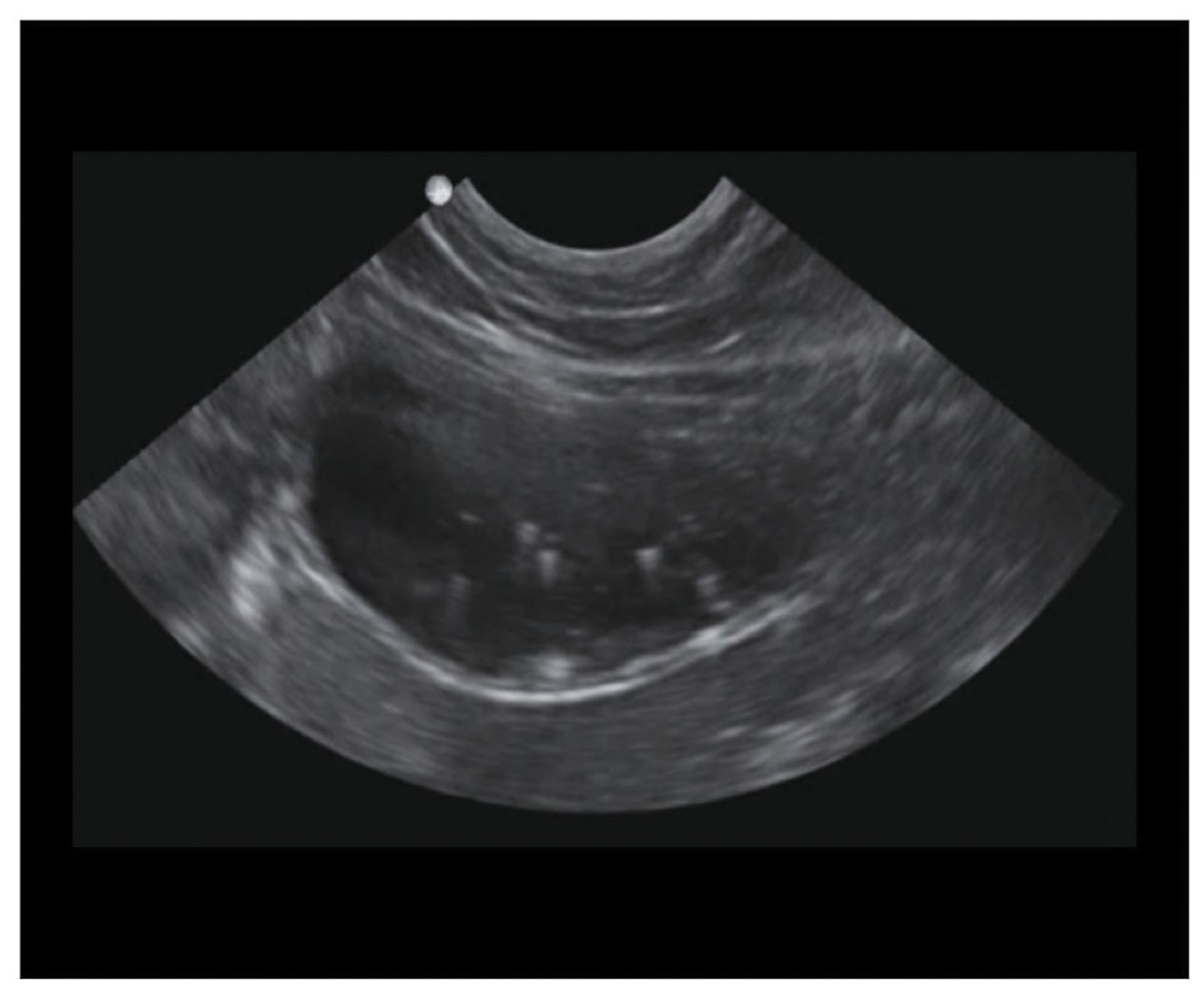
There are a number of characteristic findings on radiographs that will help your veterinarian identify the presence of pleural effusion. Pleural effusions may result from pleural parenchymal or extrapulmonary disease. The transducer is perpendicular to the ribs.
Accumulation of fluid in the pleural space. In some cases ultrasound may also be. The most commonly diagnosed cause of pleural effusion in cats is chylothorax.
The success rate is low when the effusion is loculated and septated. Four criteria have been described to differentiate ascites from pleural effusion by CT. 81 Pulmonary pathology 82 Pleural space 83 Heart and mediastinum 84 Thoracic wall.
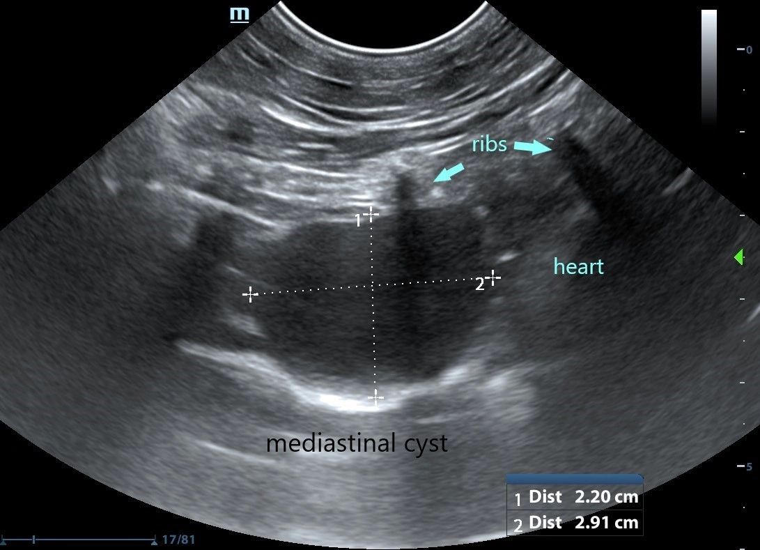
Screening for effusions can be. Unlike with a pericardial effusion in the case of accumulation of fluid in the pleural space there is no collapse of the heart walls. The caudal vena cava cvc is seen extending from the liver L to the heart H.
The type of pleural fluid withdrawn will enable your veterinarian to diagnose the cause of the pleural effusion. In the latter situations therapeutic intervention must be initiated quickly to prevent respiratory arrest. Both computed tomography CT and ultrasound US can be used to differentiate ascites from pleural effusion.
The transducer is perpendicular to the ribs. To mark the optimal site for drainage and perform the procedure if required. Fluid Scoring System TFASTÒ for the detection of pleural and pericardial effusion pneumothorax and its 4 TFASTÒ echo views and Vet BLUEÒ the veterinary brief lung ultrasound exam a regional pattern-based approach with its B-line Scoring System and its Visual Lung Language.
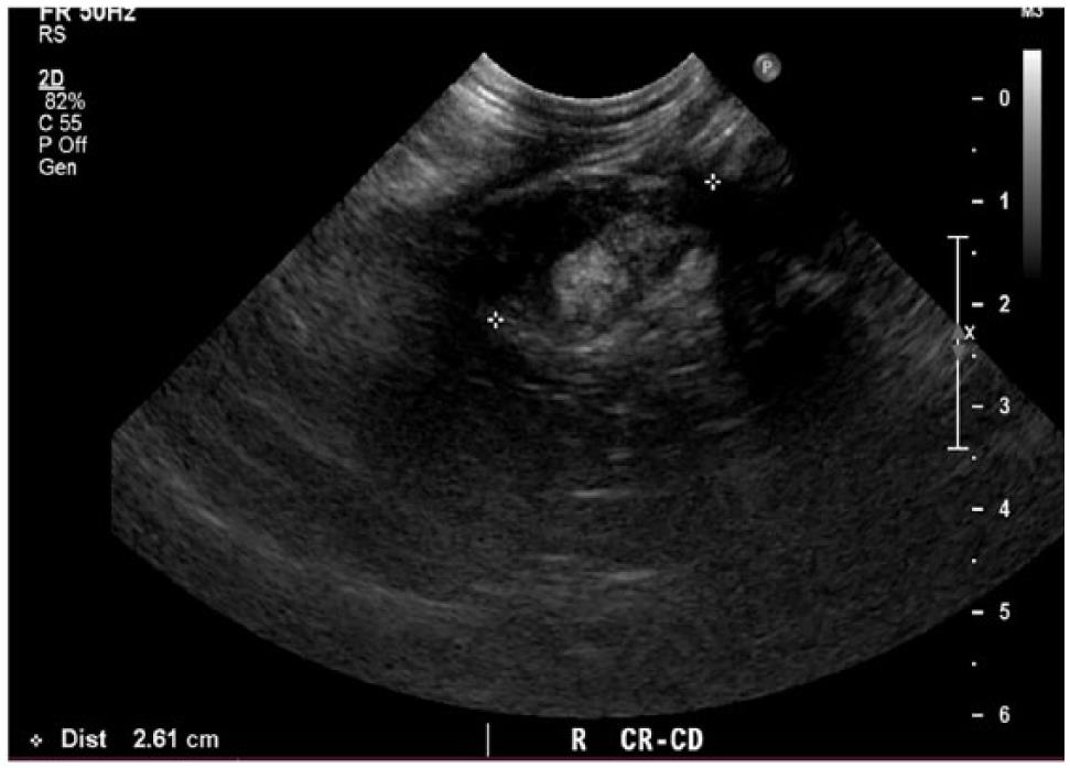
Pleural effusion is the accumulation of fluid in the pleural space resulting from disruption of the homeostatic forces responsible for the movement of pleural fluid. B Longitudinal ultrasound scan of the caudal thorax of a cat with pleural effusion. Within 458 - 287 h after the CT scan all patients were re-examined with US in the ICU.
These four signs the diaphragm sign the displaced crus sign the interface sign and the bare area sign are reliable when only one abnormal fluid collection is present. Compared to chest computed tomography CT scan pleural ultrasound has 93 sensitivity and specificity for detecting pleural effusions. Each of these 3 ultrasound formats has.
If the FAST ultrasound does reveal pleural effusion thoracentesis can be carried out. Refer to the article Pleural effusion volume ultrasound for more information. The appearance of the hematocrit sign may be observed in hemothorax with a surface layer of anechoic fluid sitting atop a settled fine echogenic sediment.
Measurement of a pleural effusion volume with point-of-care ultrasonography may be a useful tool for intensivists and is an active area of research in critical care 7. Other fluid parameters are gaining interest in veterinary patients in order to classify effusions and help determine the etiology of pleural effusion. X-ray and ultrasound imaging of the chest cavity are also very helpful in analyzing the causative factors.
This procedure removes excess fluid from the. TTE - PLAX Limited echo TTE - Anatomybasics limited echo TTE - Diagnostics Lung Trauma EFAST RenalBladder. The therapeutic intervention also provides your first diagnostic test.
Careful handling and prompt and adequate stabilisation incorporating supplemental oxygen. Limited Chest - Pleural Effusion. Ultrasonography is superior to chest radiography in detecting the presence of pleural effusions and in distinguishing pleural effusions from atelectasis or pleural thickening.
Not all veterinary practices have an ultrasound machine to perform a quick assessment in which case an x-ray can help the veterinarian evaluate the chest heart and lungs for structural abnormalities blockages and tumours. Pleural effusion is typically diagnosed by taking radiographs X-rays of the chest. In the below clip from the Sonoscape S2 you can actually see the separation of the right ventricular free wall from the pericardium in a cat.
Both the trocar and the modified Seldinger techniques can be used. Among the markers studied in cats pleural fluid lactate and total protein as well as the ratio between the pleural and serum values have higher capacity to distinguish between transudates and. In controlled settings ultrasound may detect constitutive pleural fluid can reliably detect effusions 20 mL in clinical settings and may approach the sensitivity and specificity of computed.
For those who are new to imaging around the heart with ultrasound differentiating a pericardial from a pleural effusion can be tricky particularly when the pleural effusion is circumferential around the heart. The pleural effusion volume was calculated volumetrically from the CT scan data. To determine and describe the size and site of the effusion.
Ultrasound can be used in the assessment of pleural effusion volume. Found with right congestive heart failure obstruction to lymphatic drainage by tissue adhesions in pleural space lung lobe torsion neoplasms and abdominal contents herniating. In the following article we present two cases concluding with a third case in which both types of effusion can be seen simultaneously.
Ultrasound Procedure Notes Home POCUS Exams.

Pin By Dr Abuaiad On Lymphatics Lymph Nodes Sonography Gut Health
Pleura Veterian Key

Resolution Of Nonurine Transudative Pleural Effusion In A Cat After Removal Of A Hydronephrotic Kidney In Journal Of The American Veterinary Medical Association Volume 251 Issue 1 Journals

Veterinary Echocardiography Newsletter 1 Effusions Animal Ultrasound Association
Thoracic Ultrasound

Large Secundum Asd With Right Sided Enlargement Sonography Student Heart Function Echo

Pdf Thoracic Ultrasound A Method For The Work Up In Dogs And Cats With Acute Dyspnea Semantic Scholar

Differentiating Pericardial From Pleural Effusion Animal Ultrasound Association
Front Line Ultrasound Imaging Of The Feline Urinary

Differentiating Pericardial From Pleural Effusion Animal Ultrasound Association

Resolution Of Nonurine Transudative Pleural Effusion In A Cat After Removal Of A Hydronephrotic Kidney In Journal Of The American Veterinary Medical Association Volume 251 Issue 1 Journals

Advances In Feline Cardiac Diagnostics Today S Veterinary Practice

Anticoagulent Rodenticide Toxicity Vettechlife Veterinary Vet School Veterinary Technician
Lung Ultrasound Flooding In Fulminant Pulmonary Oedema In Cats And A Comparison With Pneumonia Vet Practice Support

Search Imv Imaging

Pleural Effusion And Nt Probnp In Cats

Coughing Cats Are Not Cardiac Disease Vetgirl Veterinary Ce Blog Veterinary Cat Diseases Cardiac Disease

Echocardiography Images Echocardiography Barnard Health Care Cardiac Sonography Coarctation Of The Aorta Whipple S Disease
Spontaneous Cholecystopleural Fistula Leading To Biliothorax And Sepsis In A Cat